What is a Colposcope and How Does it Help in Detecting Cervical Cancer?
 |
| Colposcope |
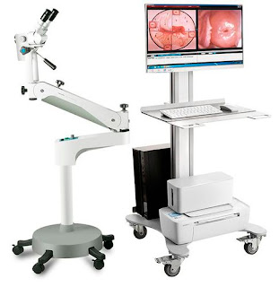
A colposcope is a medical device used to examine the cervix for any abnormalities or pre-cancerous lesions. It helps doctors detect early signs of cervical cancer or determine if further diagnostic tests are required. Here are some key things to know about colposcopy:
The Purpose and Components of a
Colposcope
A colposcope consists of a lightweight binocular microscope attached to an arm
stand. It has special lighting that illuminates, magnifies, and allows close
examination of the entire cervix and vaginal walls. During a colposcopy, a
physician places the colposcope over the vaginal opening to get a magnified
view of the cervix, which may not otherwise be visible to the naked eye.
The Colposcope
has several advantageous features compared to a normal speculum exam. Its
intense lighting technology, often using green-filtered halogen, LED or xenon
light, allows easy visualization of blood vessels and surface contours of the
cervix. Magnification ranging from 5-40X helps doctors detect any abnormal
patterns in these blood vessels or atypical bumps/lesions on the cervix.
Colposcopes also have photo documentation capabilities to record findings for
evaluating changes over time.
Reasons for a Colposcopic Exam
Colposcopy is recommended in several situations:
- Abnormal Pap smear: When a routine Pap test detects abnormal or precancerous
cells on the cervix, colposcopy can identify the exact site(s) needing further
evaluation or treatment.
- HPV infection: Women who test positive for high-risk types of human
papillomavirus (HPV), which causes majority of cervical cancers, may need colposcopy
even with normal Pap results.
- Post-procedure: Doctors perform colposcopy after treatments like cryotherapy,
LEEP or cone biopsy to check for any residual lesions requiring additional
therapy.
- Evaluation of cervical anatomy: In young women or those with a history of
multiple sexual partners, colposcopy can disclose cervical abnormalities not
detected by regular screening alone.
The Colposcopy Procedure
During the exam, a woman lies on the examination table with her knees bent and
legs supported. After inserting the speculum, the physician gently spreads it
to view the cervix and vaginal walls. They then apply a 5% vinegar/acetic acid
solution to the cervix using a cotton swab or spray.
This highlights any areas of abnormal cells, making them easier to see under
the colposcope. Normal cells temporarily turn white while precancerous cells
remain dark, irregularly bordered or raised. The doctor passes the colposcope
over the entire cervix and vagina, stopping to examine suspicious areas more closely.
They may also take cervical biopsies of any lesions using small forceps
inserted through the colposcope.
Biopsies remove tiny cell samples for laboratory analysis to definitively
diagnose conditions like cervical intraepithelial neoplasia (CIN). The
procedure itself takes 5-10 minutes and is often painless though some cramping
may occur during and after biopsies. Bleeding is minimal and the area heals
quickly. Women can resume routine activities shortly after.
Colposcopy Results and Treatment Options
After a colposcopy, possible outcomes include:
- Negative for abnormalities: The cervix appears normal and screening is
repeated according to routine guidelines.
- CIN 1: Mild changes to squamous cells on the surface of the cervix. No active
treatment needed but continued monitoring with co-testing every 1-2 years.
- CIN 2/3: Moderate to severe pre-cancerous changes detected. Outpatient
treatments like LEEP, cryotherapy or laser ablation are commonly performed to
remove the abnormal tissue.
- Invasive cancer: If an advanced stage of cervical cancer is discovered,
additional diagnostic tests and more extensive treatment involving surgery,
chemotherapy and/or radiation therapy will be required.
Get More Insights on Colpscope


Comments
Post a Comment